43 label skin layers
Label the layers of the skin Quiz - By mrumph Top Quizzes with Similar Tags. Human Muscle Anatomy 92. Countries of the World Venn Diagram III 35. European Countries Venn Diagram 18. Animals Venn Diagram 5. Corner Confusion 5. Layers of the Atmosphere 4. 5 Science Fivesomes 3. Record Label Logos 2. 5.1 Layers of the Skin - Anatomy & Physiology Skin that has four layers of cells is referred to as "thin skin.". From deep to superficial, these layers are the stratum basale, stratum spinosum, stratum granulosum, and stratum corneum. Most of the skin can be classified as thin skin. "Thick skin" is found only on the palms of the hands and the soles of the feet.
Label the Skin Quiz - PurposeGames.com This is an online quiz called Label the Skin. There is a printable worksheet available for download here so you can take the quiz with pen and paper. Your Skills & Rank. Total Points. 0. Get started! Today's Rank--0. Today 's Points. One of us! Game Points. 11. You need to get 100% to score the 11 points available.
Label skin layers
Skin Anatomy: The Layers of Skin and Their Functions The epidermis is made up of five individual layers: 2. Stratum basale: This bottom layer, also known as the basal cell layer, has column-shaped cells that push older cells toward the surface. As the cells move upward, they start to flatten and die. The layer is also made up of melanocytes (that produce a pigment that gives the skin its color ... Label the layers of the skin Diagram | Quizlet Start studying Label the layers of the skin. Learn vocabulary, terms, and more with flashcards, games, and other study tools. Solved Label the layers of the skin | Chegg.com Who are the experts? Experts are tested by Chegg as specialists in their subject area. We review their content and use your feedback to keep the quality high. 100% (38 ratings) Following are the labels of the skin from top to the bot …. View the full answer. Transcribed image text: Label the layers of the skin.
Label skin layers. A Human Body Skin-structure Quiz! - ProProfs Label B is: A. Meissner's Corpuscles. B. Pacinian Corpuscle. C. Pore. D. Free Nerve Ending. 4. Label C is: A. Dermal Papillae Layer ... These skin MCQs with answers which include multiple-choice questions on the skin will test your knowledge on the layer of protection that keeps your precious body from harm in everyday life; your skin! The skin ... Leave a Comment Cancel reply - BYJUS The skin is also responsible for maintaining our body temperature - this was apparent in victims who were subjected to the medival torture of being skinned alive. Once the skin was removed from the victim, hypothermia would set in within hours, causing the victim to freeze to death. Undoubtedly, it was also one of the most painful ways to die ... PDF Skin Diagram Labeling - New Providence School District Skin Diagram Labeling . 1. Label the diagram with the . ... You may label with a line or put the label directly onto the area described. Be as precise as possible. If you are worried about the precision of your label add a word ... U. Layers of keratinized cells V. Releases sebum . 2. Color the following structure on the diagram . Human Skin Layers - Anatomy, Functions & Fun Facts - Biology Reader Human skin layers constitute the body's outer covering that shields the internal cells, tissues, and organs against the changing environment, allergens, and pathogens. Human skin is the largest organ among the other components of the integumentary system. Besides, its immunity role, skin regulates body temperature, synthesis of vitamin-D, and ...
Solved PARTE: Assessments Sketch a vertical section of human - Chegg.com Anatomy and Physiology questions and answers. PARTE: Assessments Sketch a vertical section of human skin, using the scanning objective. Label the skin layers and a hair follicle, a sebaceous gland, and a sweat gland. You may choose to use low-power and high-power objectives to add detail to your sketch, but be sure to draw a composite drawing ... Anatomy, Skin (Integument), Epidermis - StatPearls - NCBI Bookshelf Skin is the largest organ in the body and covers the body's entire external surface. It is made up of three layers, the epidermis, dermis, and the hypodermis, all three of which vary significantly in their anatomy and function. The skin's structure is made up of an intricate network which serves as the body's initial barrier against pathogens, UV light, and chemicals, and mechanical injury. Layers of Skin: How Many, Diagram, Model, Anatomy, In Order Subcutis. The layer of skin beneath the dermis is sometimes called the subcutaneous fat, subcutis, or hypodermis layer. This layer provides insulation for your body, keeping you warm. It also ... Understanding The Different Layers Of Skin - SkinKraft Functions Of The Skin's Layers. 1. Epidermis. Epidermis is the outermost layer of your skin, making it the protective barrier which prevents the entry of harmful bacteria, viruses and other foreign substances into the deeper layers. It prevents water loss from the skin and is also responsible for its color due to the presence of melanocytes.
Skin: Cells, layers and histological features | Kenhub This article will describe the anatomy and histology of the skin. Undoubtedly, the skin is the largest organ in the human body; literally covering you from head to toe. The organ constitutes almost 8-20% of body mass and has a surface area of approximately 1.6 to 1.8 m2, in an adult. It is comprised of three major layers: epidermis, dermis and ... Label The Diagram Of The Layers Of The Skin - Learny Kids Displaying top 8 worksheets found for - Label The Diagram Of The Layers Of The Skin. Some of the worksheets for this concept are Integumentary system labeling work answers, Title skin structure, Integumentary system work basic skin structure, Label the skin anatomy diagram answers, Name your skin, Section through skin, Inside earth work, Anatomy physiology. Label the Skin Layers - Printable - purposegames.com This is a free printable worksheet in PDF format and holds a printable version of the quiz Label the Skin Layers. By printing out this quiz and taking it with pen and paper creates for a good variation to only playing it online. This printable worksheet of Label the Skin Layers is tagged. Click on the tags below to find other worksheets in the ... Epidermis (Outer Layer of Skin): Layers, Function & Structure While the epidermis is the thinnest layer of skin, the dermis is the thickest layer of skin. The dermis contains collagen and elastin, which help make it so thick and supportive of your skin's overall structure. All of your connective tissues, nerve endings, sweat glands, oil glands and hair follicles exist in the dermis as well as the ...
ANAT2241 Connective Tissue Components - Embryology
The Three Layers of Skin and Their Functions | FLDSCC 3. The Hypodermis. The hypodermis is made of subcutaneous (under the skin) fats, connective tissues, blood vessels, and nerve cells. It's the layer of skin where fat is deposited and stored. The blood vessels in the hypodermis are bigger and connect to the rest of your body. Stored fat helps regulate body tissue and cushion your body's ...

Sectioned Sebaceous - Human Anatomy - GUWS Medical
Human Biology fig. 1.24 - Layers of the skin - English labels Layers of the skin. The inner layer of the skin is the dermis, and the outer layer is the epidermis. The epidermis can be specified further in the stratum corneum, stratum lucidum, stratum gransulosum, stratum spinosum and stratum basale. English labels. From 'Human Biology' by D. Wilkin and J. Brainard . You are free to use this item if ...

Integumentary System - Histology - Embryology
Answered: 6. Label the layers of the skin to… | bartleby 6. Label the layers of the skin to their correct location by clicking and dragging the labels to the micrograph image. (Some labels may not. be used.) CopyrigheThe Mr Compas Pmisso d noday Dermal papilla Stratum lucidum Stratum basale Hypodermis Stratum corneum Stratum spinosum Dermis Stratum granulosum The MoGrow H Compares e.Denns Sete ...

Pin on Histology - Skin
Creative 20 Label Skin Diagram Worksheet - Worksheet Template Ideas Whether your child needs a label skin diagram worksheet or is interested in learning more about the label skin diagram worksheet, our free worksheets and printable activities cover all the educational bases. Each worksheet was created by a professional educator, so you know your child will learn critical age-appropriate facts and concepts.

Classification of Skin Burns | Doctor Stock
Label Skin Diagram Printout - EnchantedLearning.com Read the definitions, then label the skin anatomy diagram below. blood vessels - Tubes that carry blood as it circulates. Arteries bring oxygenated blood from the heart and lungs; veins return oxygen-depleted blood back to the heart and lungs. dermis - (also called the cutis) the layer of the skin just beneath the epidermis.
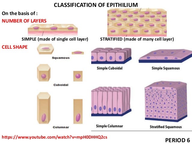
Tissues Class 9 ppt
Layers of the skin: label Diagram | Quizlet Start studying Layers of the skin: label. Learn vocabulary, terms, and more with flashcards, games, and other study tools.
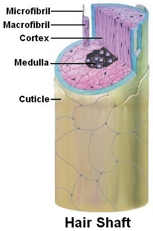
Basic Hair Structure - Hair Follicle and Hair Shaft Function
Integumentary System Diagram Labeled The integumentary system is an organ system consisting of the skin, hair, nails, and exocrine glands. The skin is only a few millimeters thick yet is by far the largest organ in the body. The epidermis is the most superficial layer of the skin that covers almost the entire body surface. The epidermis rests upon and protects the deeper and ...

Life as I see it: THICK SKIN IS SOMETIMES VITAL
Skin Labeling | Biology Game | Turtle Diary Identify and label figures in Turtle Diary's interactive online game, Skin Labeling! Drag the given words to the correct blanks to complete the labeling! Skin Labeling - Biology Game. Bly8337. 35,274 Plays Grade K - 5 (650) Skin Labeling.
Post a Comment for "43 label skin layers"