40 dissected cow eye labeled
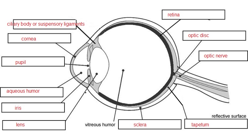
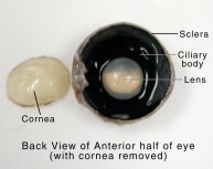
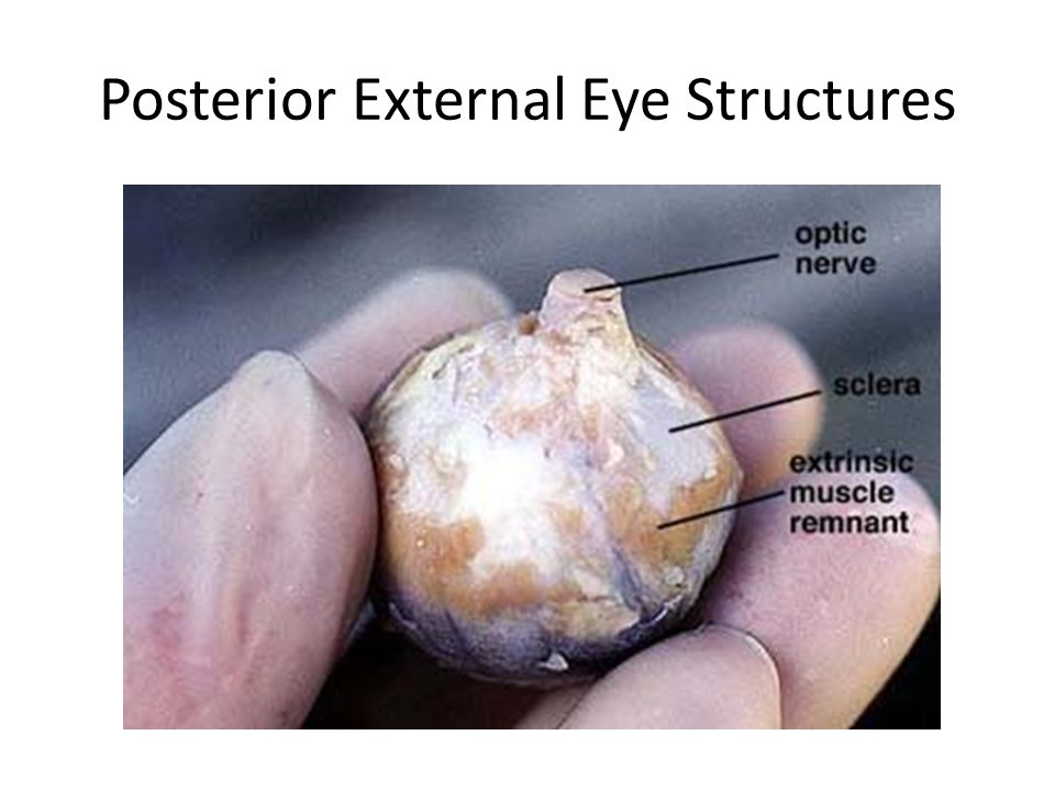
eye diagram labeled Biology: Sheep Eye Dissection albertsscientist4com.blogspot.com. eye sheep dissection external cornea dissected optic nerve structures biology left. LAS Assignment 7 - Eye Enucleation cmapsconverted.ihmc.us. eye cow anatomy human eyes dissection enucleation lucidum tapetum ihmc science rid. Eye anatomy diagram quiz basic game purposegames. PDF Cow Eye Dissection Guide - Central Bucks School District slide out of the eye. You may need to gently tease it loose from the inside of the eye. 3. The eyeball should now be separated into an anterior and a posterior half. Turn the anterior portion of the eyeball over on the dissecting tray so External view of the cow eye
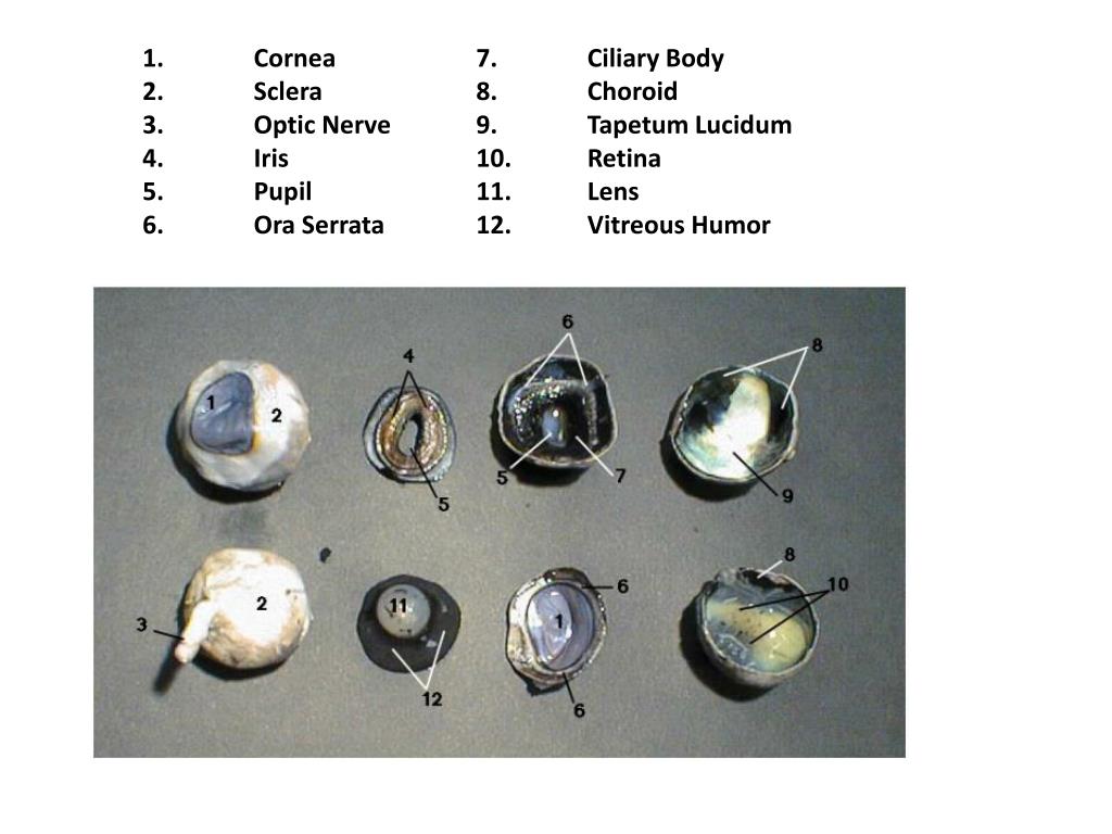
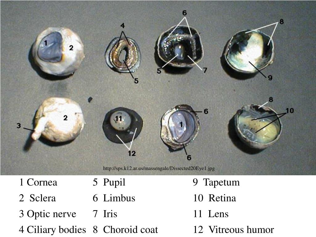
Solved PART C: Assessments Complete the following: FIGURE - Chegg PART C: Assessments Complete the following: FIGURE 35.14 Partial frontal cut of dissected cow eye. Label the internal structures using the list provided. 3 Ciliary body Lens Retina Sclera Tapetum fibrosum of choroid)

Dissected cow eye labeled
Cow Eye Dissection & Labeling - YouTube About Press Copyright Contact us Creators Advertise Developers Terms Privacy Policy & Safety How YouTube works Test new features Press Copyright Contact us Creators ... Lab 10—Labeled Cow Eye Lab 10—Labeled Cow Eye BIO 137 Virtual Lab 10 The Cow Eye (Labeled) Return to: Lab 10 PageBIO 137 Main Page Be sure to practice using the unlabeled images. Coronal section, anterior view (lens and vitreous humor displaced) Sagittal section This page created and maintained by Udo M. Savalli. Last updated April 18, 2006. Cow Eye Dissection & Anatomy Project | HST Learning Center Cow Eye Dissection: Internal Anatomy 1. Place the cow's eye on a dissecting tray. The eye most likely has a thick covering of fat and muscle tissue. Carefully cut away the fat and the muscle. As you get closer to the actual eyeball, you may notice muscles that are attached directly to the sclera and along the optic nerve.
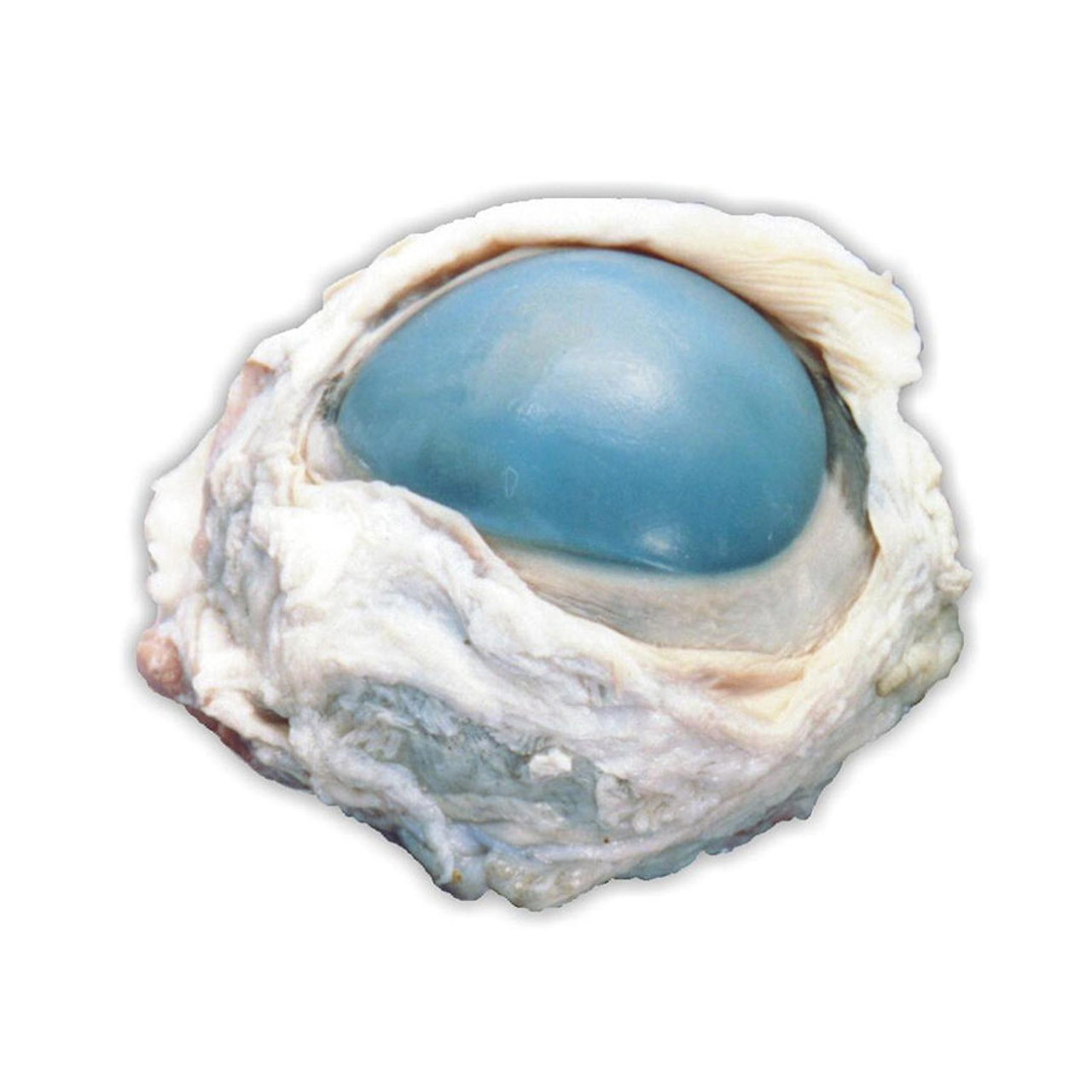
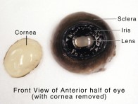
Dissected cow eye labeled. Cow's Eye Dissection - Eye diagram - Exploratorium A cow's iris is brown. Human irises come in many colors, including brown, blue, green, and gray. A clear fluid that helps the cornea keep its rounded shape. The pupil is the dark circle in the center of your iris. It's a hole that lets light into the inner eye. Your pupil is round. A cow's pupil is oval. PDF COW'S EYE dissection - Exploratorium the covering over the front of the eye (the cornea). When the cow was alive, the cornea was clear. In your cow's eye, the cornea may be cloudy. You may be able to look through the cornea and see the iris, the colored part of the eye, and the pupil, the dark oval in the middle of the iris. Cut away the fat and muscle. Here's what you do: 1 2 › 43785083 › The_Hidden_DimensionThe Hidden Dimension - Edward Hall - Academia.edu Enter the email address you signed up with and we'll email you a reset link. neilpatel.com › blogNeil Patel's Digital Marketing Blog By clicking the button below, you consent for NP Digital and partners to use automated technology, including pre-recorded messages, cell phones and texts, and email to contact you at the number and email address provided.
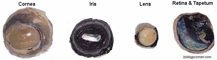
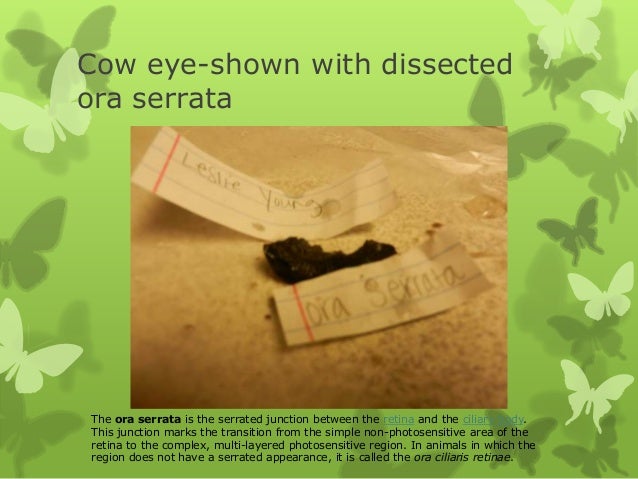
Cow eye - dissection and label Cow eye - dissection and label 1. Cow eye - Dissection and Label By Leslie Young Section 63 2. Cow eye shown with labeled cornea. The cornea is the transparent front part of the eye that covers the iris, pupil,... 3. Cow eye-shown with labeled Sclera The sclera (from the Greek skleros, meaning hard ... View 26 Cow Eye Dissection Labeled Diagram - withoutcolorbox We Have got 15 picture about Cow Eye Dissection Labeled Diagram images, photos, pictures, backgrounds, and more. In such page, we additionally have number of images out there. Such as png, jpg, animated gifs, pic art, symbol, blackandwhite, images, etc. If you're searching for Cow Eye Dissection Labeled Diagram topic, you have visit the ideal ... Cow Eye Dissection - The Biology Corner 1. Examine the outside of the eye. You should be able to find the sclera, or the whites of the eye. This tough, outer covering of the eyeball has fat and muscle attached to it 2. Locate the covering over the front of the eye, the cornea. When the cow was alive, the cornea was clear. In your cow's eye, the cornea may be cloudy or blue in color. 2. brain parts labelled diagram brain parts labelled diagram. Brain shark labeled nervous cranial dogfish system anatomy spinal dissected cavity nerves organs primitive human non animals very sense cord. Sheep brain labeled. Eye anatomy cow dissection labeled sheep section parts human lab sagittal lens eyes physiology quizlet internal student animal system choroid. Smith, K ...
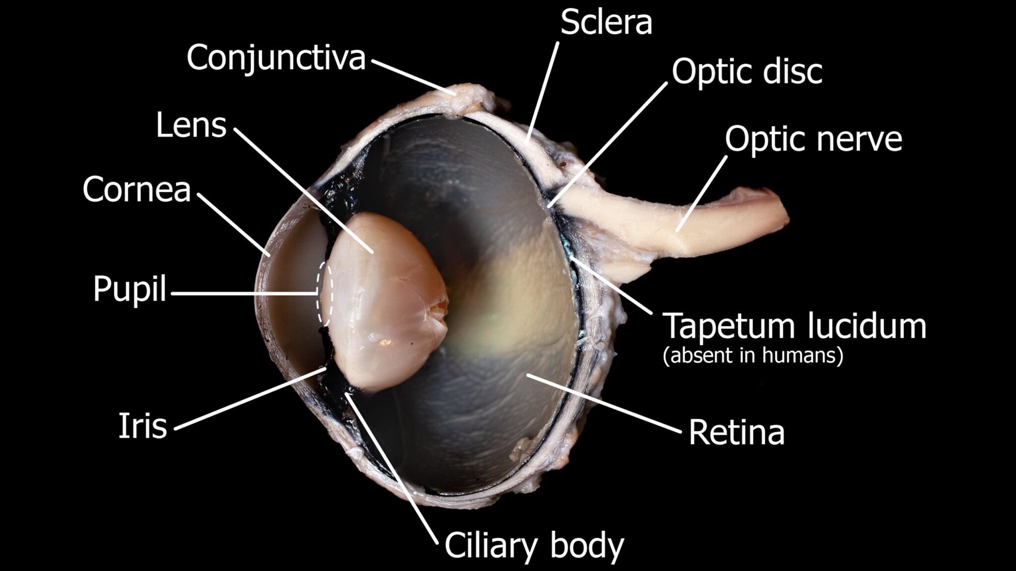
nlp.stanford.edu › ~lmthang › morphoNLMThe Stanford Natural Language Processing Group ' '' ''' - -- --- ---- ----- ----- ----- ----- ----- ----- ----- ----- ----- ----- ----- ----- ----- ----- ----- ----- ----- ----- ----- ----- ----- ----- ----- ----- ----- ----- ----- ----- ----- ----- ----- ----- ----- ----- ----- ----- ----- ----- ----- ----- ----- ----- ----- ----- ----- ----- ----- ----- ----- ----- ----- ----- ----- ----- ----- ----- ----- Describe the Lens of the Dissected Cow Eye Learn how to dissect a cows eye in your classroom. Cow Eye Quiz Dissection 101 Click Ppt Download Use the structures listed in question 2 and label the diagram.. Sends messages from the eye to the brain. During this activity you will dissect a cow eye. Outer lens that lets light into the eye. Up to 24 cash back 2. Parts and Functions - Dissecting A COW EYE Iris and Pupil. The iris is a muscle that controls how much light goes into the eye and suspended between the cornea and lens. As we have seen in the dissection a cow's iris is brown. The pupil is the dark circle that's in the center of the iris and it lets light into the inner eye. A&P Lab II - Dissected Cow Eye Flashcards | Quizlet adipose (fatty) cushion identify the structure labeled "1" choroid identify the structure ciliary body identify the structure labeled "6" conjunctiva cornea identify the structure labeled "2" iris identify the structure lens identify the structure labeled "7" extrinsic eye muscle identify the structure labeled "5" optic nerve
Fresh cow eye dissection (Part 3): Iris and retina
Lab 11 eye dissection - Human Biology Lab 11 Cow eye... View Lab Report - Lab 11 eye dissection from BSC 2020C at Florida State College at Jacksonville. Human Biology Lab 11 Cow eye dissection Kelly Kopit-Bond Purpose: The main purpose of this lab is to
Cow Eye Dissection
huggingface.co › hf-internal-testing › tiny-randomHugging Face – The AI community building the future. ・Carolina熱「 ・Homer熱」 ・tournament熱、 ・conflict熱・ ・floor熱ヲ ・toward熱ァ ・singles熱ィ ・couple熱ゥ ・except熱ェ ・formation熱ォ ・reference熱ャ ・Scottish熱ュ ・develop熱ョ ・setting熱ッ ・speech熱ー ・territory熱ア ・worldwide熱イ ・medical熱ウ ・1987熱エ ・trying熱オ ...
SCB209 - Lab3 - Natural Sciences Open Educational Resources
Solved Activity 3: Dissecting the Mammalian Eye 1. Identify - Chegg Question: Activity 3: Dissecting the Mammalian Eye 1. Identify the labeled structures in the accompanying photographs of a dissected cow eye a. b. c. d. e. Activity 4: Performing Visual Tests 1. Where specifically is the blind spot located? 2. What does 20/100 vision mean? 3. Define astigmatism. 4. What causes color blindness? 5.

Science - *Zakiabizk*
Cow Eye Dissection Worksheet | Cow eyes, Dissection, Frog ... - Pinterest Eye Definition The first line of protection of the eyes is provided by the lids, which prevent access of foreign bodies and assist in the lubrication of the corneal surface. Lid closure and opening are accomplished by the orbicularis oculi and levator palpebri muscles; the orbicularis oculi operates on both lids, bringing their margins into close apposition in the act of lid closure.

Carolina® Eye Dissection Mat
Cow Eye Dissection Teaching Resources | Teachers Pay Teachers The guide includes step-by-step instructions and labeled diagrams that will lead students through the external anatomy of the eye, followed by dissection of the internal structures. ... Next, a cow eye is dissected and the pertinent structures are identified including: tear gland, cornea, sclera, iris, pupil, optic nerve, lens, retina, choroid ...
PPT - Sheep Eye Dissection PowerPoint Presentation, free ...
Cow Eye Dissection Quiz - PurposeGames.com cow eye, dissection Remaining 0. Correct 0. Wrong 0. Press play! 0%. 0:00.0. Quit. Again. This game is part of a tournament. You need to be a group member to play the tournament. Join group, and play Just play. Your Scorecard. The scorecard of a champion. Score . 0 % Time . 0:00.0. Place # 0.
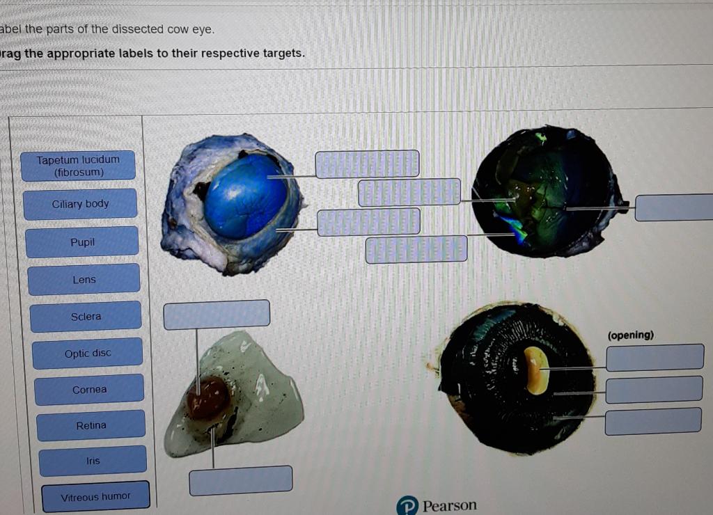
abel the parts of the dissected cow eye. rag the | Chegg.com
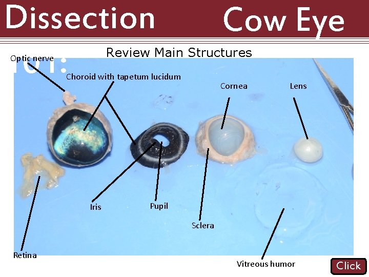
Cow Eye Dissection | Carolina.com Cow Eye External Anatomy. Obtain a preserved cow eye, and then place it on your dissecting tray. Examine the external characteristics of the eye. Use the image below and identify: Cornea—at front of the eye; allows light to enter the eye; Optic nerve—at back of the eye; transmits nerve impulses to the brain; Fat; Muscle
_BIOLOGY 20 Lab ManualV2A
› 2022 › 08MoA - Open (NOT Ukraine) Thread 2022-126 Aug 11, 2022 · 2-fer: abortion and FB giving up data on users. This is the data facebook gave police to prosecute a teenager for abortion. According to court records, Celeste Burgess, 17, and her mother, Jessica Burgess, bought medication called Pregnot designed to end pregnancy.
Cow Eye Dissection Welcome to Cow Eye Dissection. This ...
Cow Eye Dissection & Parts of the Eye Diagram | Quizlet A ring of muscle tissue that forms the colored portion of the eye; controls the size of the pupil opening. retina. Located in the back of the eye, contains the rods and cones. pupil. The opening in the center of the iris through which light enters the eye. lens.

WMU Psychology Department: Lisa Baker
Lab HW 11 Cow Eye.pdf - Bio 210 - Lab HW 11 (6 Points)... View Lab HW 11 Cow Eye.pdf from 806 177 at Waukesha County Technical College. Bio 210 - Lab HW 11 (6 Points) Name: Teresa Nguyen Part I. Label structures from your dissected cow eye. Part II. Draw

Eye Anatomy Handout - Docsity
cow leg anatomy Dissected Cow Eye Clearly Showing How Eye Can Be Understood And How eye cow lens anatomy dissected dissection labeled eyes human camera vet cat clearly understood showing sagittal section muscle open Anatomical Model Of Cow Leg At 1stdibs 1stdibs.com anatomical Body Parts Of The Cute Cartoon Farm Cow.
PPT - COW EYE DISSECTION PowerPoint Presentation, free ...
Cow Eye Dissection Guide - Google Slides Cow Eye. Use the point of a scissors or a scalpel to make an incision through the layers of the eye capsule (similar to figure 1); there are three layers from the exterior: sclera, whitish/grey,...
Dissection 101 Reasons to Use the Dissection Video
PDF Cow Eye Dissection: Examining Structure and Function - Woodstown The eyes of cows are structurally and functionally similar to the eyes of humans. During this activity, you will dissect a cow eye. You will observe several important features of the eye and develop your understanding of how each part functions to make vision possible. Materials • Preserved Cow Eye • Scalpel or Scissors • Forceps

Cow Eye Dissection - Perkins School for the Blind
Cow Eye Dissection & Anatomy Project | HST Learning Center Cow Eye Dissection: Internal Anatomy 1. Place the cow's eye on a dissecting tray. The eye most likely has a thick covering of fat and muscle tissue. Carefully cut away the fat and the muscle. As you get closer to the actual eyeball, you may notice muscles that are attached directly to the sclera and along the optic nerve.

Lab 10—Labeled Cow Eye
Lab 10—Labeled Cow Eye Lab 10—Labeled Cow Eye BIO 137 Virtual Lab 10 The Cow Eye (Labeled) Return to: Lab 10 PageBIO 137 Main Page Be sure to practice using the unlabeled images. Coronal section, anterior view (lens and vitreous humor displaced) Sagittal section This page created and maintained by Udo M. Savalli. Last updated April 18, 2006.
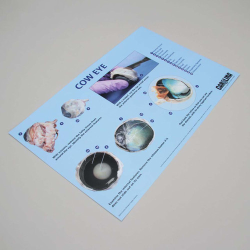
Cow eye dissection mat 28cm x 43cm
Cow Eye Dissection & Labeling - YouTube About Press Copyright Contact us Creators Advertise Developers Terms Privacy Policy & Safety How YouTube works Test new features Press Copyright Contact us Creators ...
![Anatomy of the eye [17]. | Download Scientific Diagram](https://www.researchgate.net/profile/Ali-Taqi-8/publication/315769958/figure/fig1/AS:533481529593856@1504203316282/Anatomy-of-the-eye-17_Q640.jpg)
Anatomy of the eye [17]. | Download Scientific Diagram

Sheep Eye Disection | Mohtadi Alkhaliq
Cow Eye Dissection & Anatomy Project | HST Learning Center

DISSECTING A COW'S EYE
Cow Eye Dissection

Cow Eye Dissection - Perkins School for the Blind

Cow's Eye Dissection - Eye diagram

COW EYE DISSECTION - NANDINI SONI

Cow Eye Dissection Diagram | Quizlet
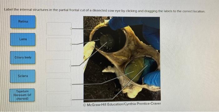
Solved Label the internal structures in the partial frontal ...

Cow Eye Dissection Guide - Google Slides
Cow Eye for Dissection Specimen
Sheep Eye Dissection. - ppt video online download
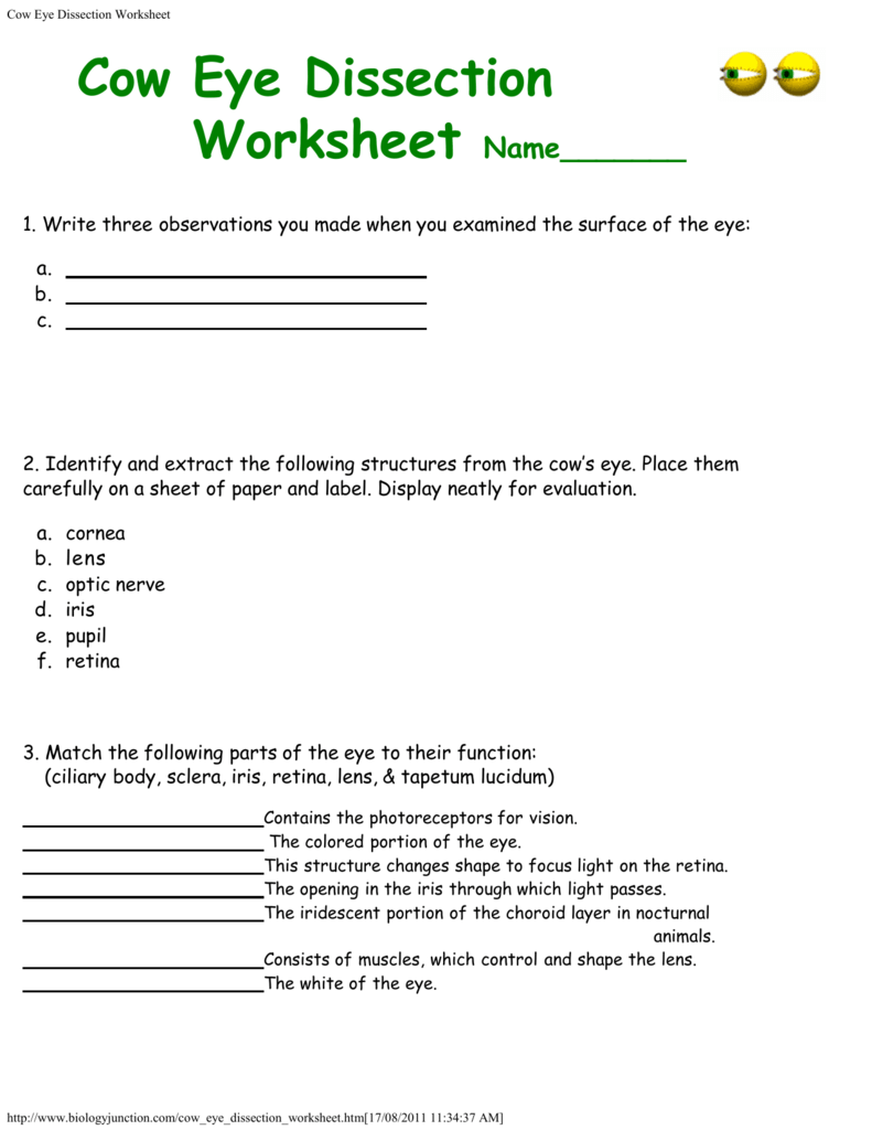
Cow Eye Dissection Worksheet
Cow Eye Dissection & Anatomy Project | HST Learning Center

Funventure: Cow Eye Dissection, Discovery Outpost, Medicine ...

Cow Eye Dissection | Carolina.com
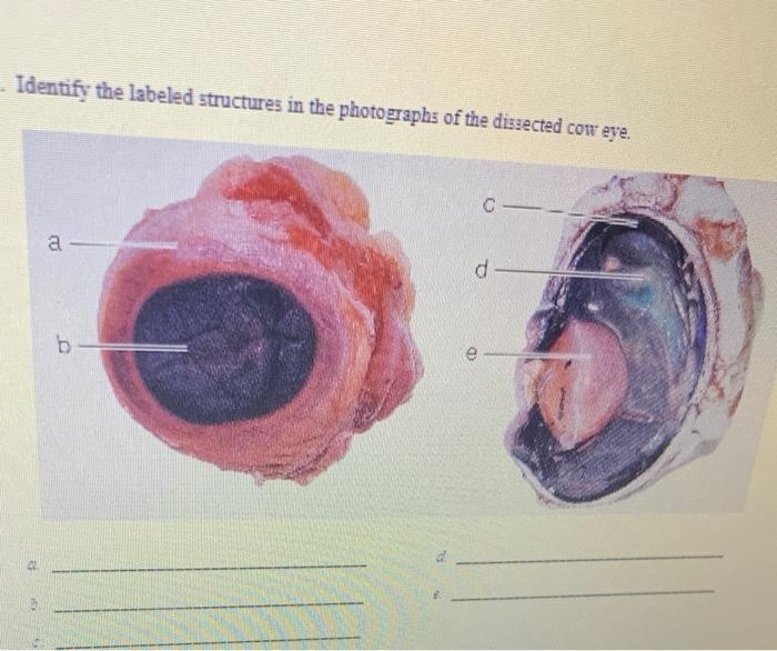
Solved Identify the labeled structures in the photographs of ...
Cow eye – dissection and label

Cow Eye Labeling Quiz
Hamburg CSD - 5th Grade Cow Eyes

Cow Eye Dissection | Carolina.com

Cow Eye Dissection Quiz

Cow Eye Dissection Post Lab - Aidan - Eye Dissection Post Lab ...
Post a Comment for "40 dissected cow eye labeled"