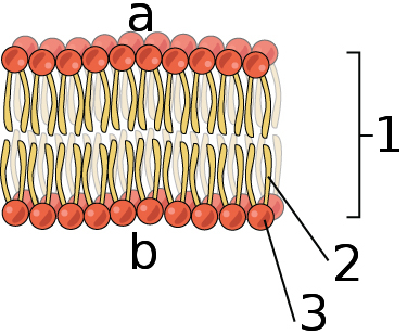
39 draw and label a phospholipid bilayer
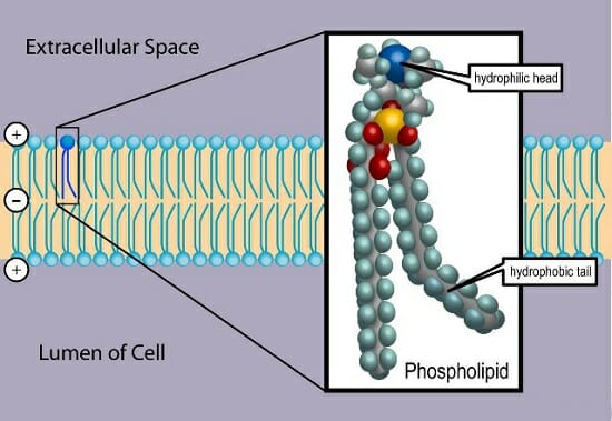
Campbell Biology, 12th Edition [12 ed.] 9780135988046 Verkko35. On these diagrams of plant and animal cells, label each organelle and give a brief statement of its function. NEW! Active Reading Guides support students in actively reading their biology text. Students can download the worksheets from the Study Area in Mastering Biology. leonhardt-rechtsanwaelte.de › diagram-of-a-cellThe structure and components of a human cell are given below ... This figure depicts the lipid bilayer and the structure of a phospholipid: Functions of Phospholipids modified stableford scoring with handicaps The fluid mosaic model describes cell membranes as ‘mosaics’ because: The scattered pattern produced by the proteins within the phospholipid bilayer looks somewhat like a mosaic when viewed from ...
wou.edu › chemistry › chapter-11-introduction-majorCH103 – Chapter 8: The Major Macromolecules – Chemistry The simplest of these structures are micelles, spherical assemblies containing a hydrophobic interior of phospholipid tails and an outer surface of polar head groups. Larger and more complex structures are created from lipid-bilayer sheets, or unit membranes, which are large, two-dimensional assemblies of phospholipids congregated tail to tail.
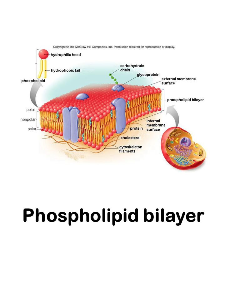
Draw and label a phospholipid bilayer
BioChem Ch 10 Flashcards | Quizlet VerkkoStudy with Quizlet and memorize flashcards containing terms like Classify the following molecules as lipids (or lipid components), carbohydrates, or nucleic acids., Hydrogenation of a monounsaturated fatty acid yields a saturated fatty acid. Oleic acid, CH3(CH2)7CH=CH(CH2)7CO2H is a monounsaturated fatty acid. Predict the product … Construction of the Cell Membrane - Wisc-Online OER VerkkoLearners read and listen to the pronunciation of hundreds of medical terms that are arranged in a "jukebox." The terms are listed alphabetically and according to the following categories: aerobic and anaerobic microorganisms, blood bank, coagulation, fungi microorganisms, hematology, protozoa, and urinalysis. Cambridge International AS & A Level Biology Coursebook … Verkkosections of Practical Activity 7.1 before answering the question below, which is relevant to this chapter. Figures 1.8a and b show examples of good drawing and labelling technique based on Figures 1.6 and 1.7. Note that it is acceptable to draw only a representative portion of the cell contents of Figure 1.7, but add a label explaining this.-R
Draw and label a phospholipid bilayer. › 44090147 › Cambridge(PDF) Cambridge International AS and A Level Biology ... BIO1: Maintaining a Balance 1. Most organisms are active in a limited temperature range IDENTIFY THE ROLE OF ENZYMES IN METABOLISM, DESCRIBE THEIR CHEMICAL COMPOSITION AND USE A SIMPLE MODEL TO DESCRIBE THEIR SPECIFICITY ON SUBSTRATES Biochemistry Final Review Questions Flashcards | Quizlet VerkkoDraw the structure of the product from each reaction as it would exist at pH 7. Include the appropriate hydrogen atoms. Draw the structure of the group attached to −SCoA. ... the average cross‑sectional areas of a 50,000 Da protein and a 750 Da phospholipid in a bilayer. ... Label the enzymes and compounds of the carnitine shuttle system. CH103 – Chapter 8: The Major Macromolecules – Chemistry VerkkoProteins are very large molecules containing many amino acid residues linked together in very specific order. Proteins range in size from 50 amino acids in length to the largest known protein containing 33,423 amino acids. Macromolecules with fewer than 50 amino acids are known as peptides.. Figure 11.4 Peptides and Proteins are macromolecules … dokumen.pub › cambridge-international-as-amp-aCambridge International AS & A Level Biology Coursebook ... sections of Practical Activity 7.1 before answering the question below, which is relevant to this chapter. Figures 1.8a and b show examples of good drawing and labelling technique based on Figures 1.6 and 1.7. Note that it is acceptable to draw only a representative portion of the cell contents of Figure 1.7, but add a label explaining this.-R
Academia.edu - Cambridge International AS and A Level Biology ... VerkkoBIO1: Maintaining a Balance 1. Most organisms are active in a limited temperature range IDENTIFY THE ROLE OF ENZYMES IN METABOLISM, DESCRIBE THEIR CHEMICAL COMPOSITION AND USE A SIMPLE MODEL TO DESCRIBE THEIR SPECIFICITY ON … 2.4.1 The Structure of Cell Membranes - Save My Exams VerkkoThe scattered pattern produced by the proteins within the phospholipid bilayer looks somewhat like a mosaic when viewed from above; ... You must know how to draw and label the fluid mosaic model, ... An example of the diagram you could draw. Test Yourself Next Topic. 1. Biological Molecules. 1.1 Biological Molecules: ... › figure › Radioactive-LabelingRadioactive Labeling of Galactolipids in Chloroplast ... Download Table | Radioactive Labeling of Galactolipids in Chloroplast Membranes and lntact Seedlings from publication: Isolation and Characterization of an Arabidopsis Mutant Deficient in the ... quizlet.com › 505043992 › biochem-ch-10-flash-cardsBioChem Ch 10 Flashcards | Quizlet Study with Quizlet and memorize flashcards containing terms like Classify the following molecules as lipids (or lipid components), carbohydrates, or nucleic acids., Hydrogenation of a monounsaturated fatty acid yields a saturated fatty acid. Oleic acid, CH3(CH2)7CH=CH(CH2)7CO2H is a monounsaturated fatty acid. Predict the product of its hydrogenation:, Draw the fat molecule whose components ...
Join LiveJournal VerkkoPassword requirements: 6 to 30 characters long; ASCII characters only (characters found on a standard US keyboard); must contain at least 4 different symbols; (a) Steady-state emission spectra (λ ex = 358 nm) for Prodan in … VerkkoThe temperature was maintained at 10 @BULLET C. from publication: Probing Ethanol-Induced Phospholipid Phase Transitions by the Polarity Sensitive Fluorescence Probes Prodan and Patman ... › learn › natural-scienceConstruction of the Cell Membrane - Wisc-Online OER Learners read and listen to the pronunciation of hundreds of medical terms that are arranged in a "jukebox." The terms are listed alphabetically and according to the following categories: aerobic and anaerobic microorganisms, blood bank, coagulation, fungi microorganisms, hematology, protozoa, and urinalysis. Cambridge International AS & A Level Biology Coursebook … Verkkosections of Practical Activity 7.1 before answering the question below, which is relevant to this chapter. Figures 1.8a and b show examples of good drawing and labelling technique based on Figures 1.6 and 1.7. Note that it is acceptable to draw only a representative portion of the cell contents of Figure 1.7, but add a label explaining this.-R
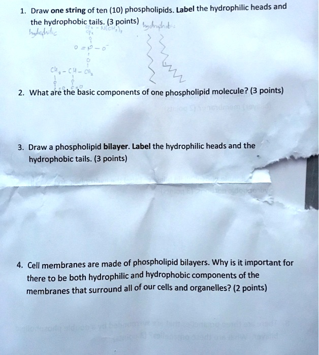
SOLVED: Draw one string of ten (10) phospholipids. Label the ...
Construction of the Cell Membrane - Wisc-Online OER VerkkoLearners read and listen to the pronunciation of hundreds of medical terms that are arranged in a "jukebox." The terms are listed alphabetically and according to the following categories: aerobic and anaerobic microorganisms, blood bank, coagulation, fungi microorganisms, hematology, protozoa, and urinalysis.
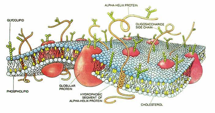
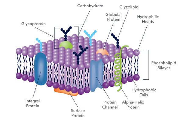
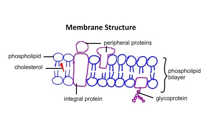
Diagram of a cell membrane with labels
BioChem Ch 10 Flashcards | Quizlet VerkkoStudy with Quizlet and memorize flashcards containing terms like Classify the following molecules as lipids (or lipid components), carbohydrates, or nucleic acids., Hydrogenation of a monounsaturated fatty acid yields a saturated fatty acid. Oleic acid, CH3(CH2)7CH=CH(CH2)7CO2H is a monounsaturated fatty acid. Predict the product …

Biology Notes for A level: #27 Summary of Cell membrane

Diagram of the cell membrane | Download Scientific Diagram

Phospholipid Bilayer Diagram | Quizlet
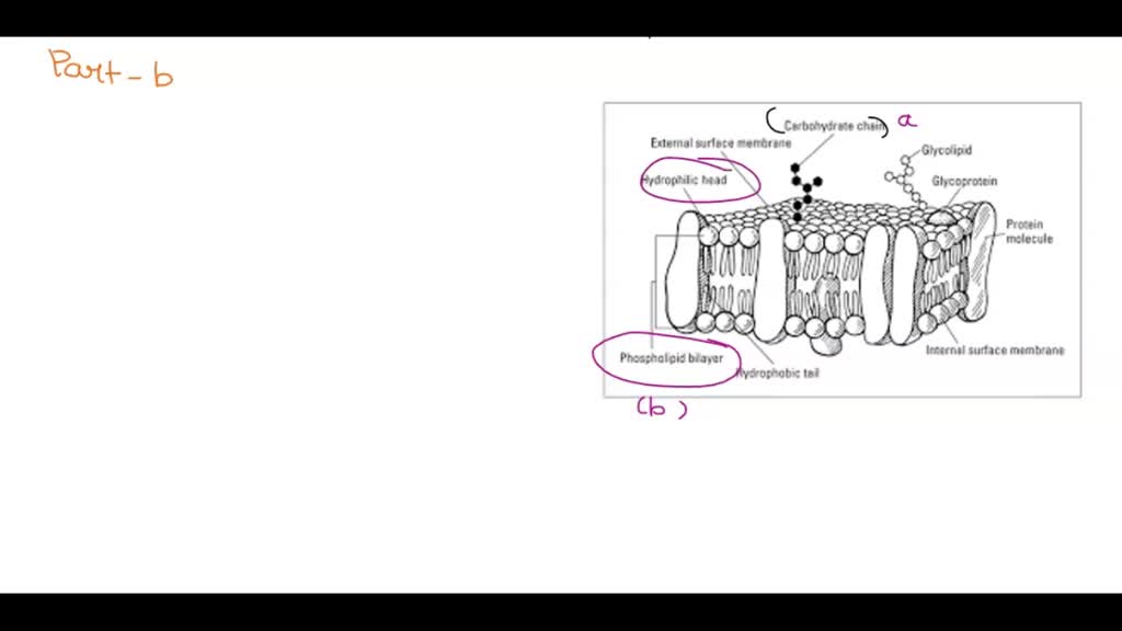
Part 1: Why is the model of the cell membrane referred to as? fluid, mosaic, Part 2: On the diagram of membrane below; label the following parts: carbohydrate; phospholipid; cholesterol and ...
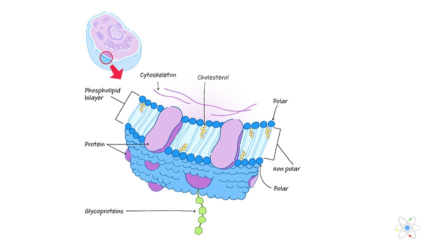
Plasma Membrane: Definition, Structure & Function (with ...

2.4.1 Draw and label a diagram to show the structure of ...

The Phospholipid molecule Hydrophilic – “water loving ...

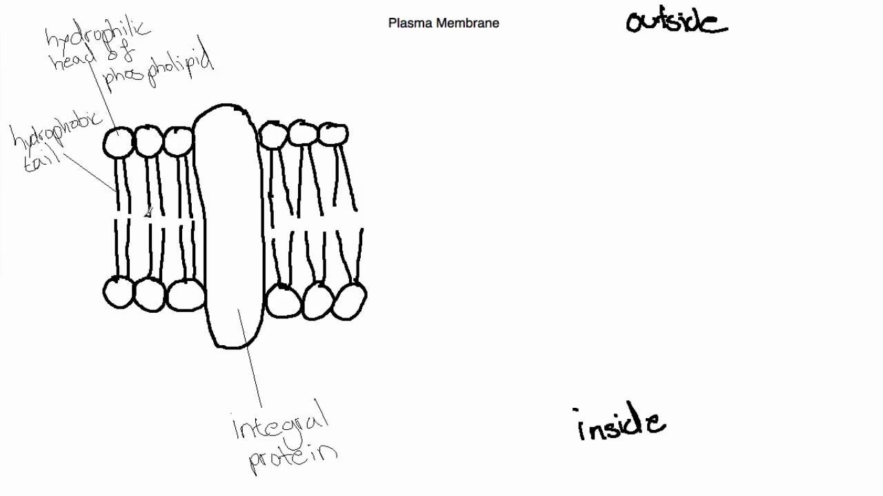
The Cell Membrane Write. Cell Membrane The membrane of the ...

Fluid mosaic model: cell membranes article (article) | Khan ...

Drawing Fluid Mosaic Model Of Plasma Membrane in Easy Way | Drawing Cell Membrane
File:0302 Phospholipid Bilayer labeled.jpg - Wikimedia Commons
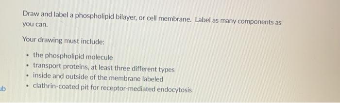
Solved Draw and label a phospholipid bilayer, or cell | Chegg.com
Plasma Membrane Markers: Novus Biologicals
IB Biology Topic 2.4.1 Draw and Label the Plasma Membrane

Label the Phospholipid Bilayer Diagram | Quizlet

3.5: Lipid Molecules - Phospholipids - Biology LibreTexts

Label the Cell Membrane Diagram | Quizlet

Describing the Structure and Nature of a Phospholipid
Phospholipid: Definition, Structure, Function | Biology ...

Drawing the fluid-mosaic model (2016) IB Biology

IB Biology 2.4.1: Drawing a plasma membrane - YouTube

How to Draw a Phospholipid Bilayer
DP Biology: Membrane Structure
1.) On the following diagram of a phospholipid bilayer; label the hydrophobic ad hydrophilic portions of each phospholipid molecule:, 2.) What molecule is found in the plasma membrane of animals but ...

Label Cell Membrane Diagram | Quizlet

Phospholipid Bilayer | CK-12 Foundation

Phospholipids (1.2.3) | AQA A Level Biology Revision Notes ...

1,765 Phospholipids Images, Stock Photos & Vectors | Shutterstock
Topic 1.3 Membrane Structure - AMAZING WORLD OF SCIENCE WITH ...

Solved Activities MC-N-CH, CH, CH, 0 Membrane components A ...
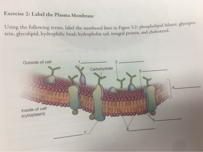
Solved Exercise 2: Label the Plasma Membrane Using the ...
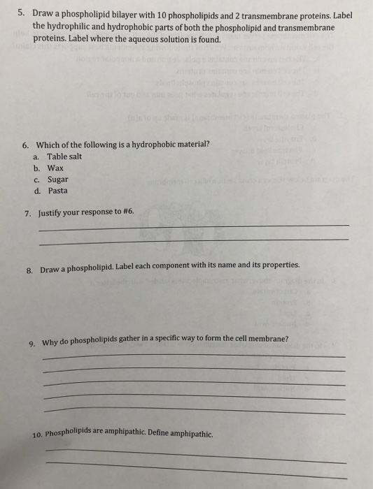
Solved Draw a phospholipid bilayer with 10 phospholipids and ...

Fluid mosaic model: cell membranes article (article) | Khan ...

Cell Membrane Structure - Exchange and Transport Ep 1 - Zoë ...
Phospholipid bilayer. Prepare to draw key components to help ...
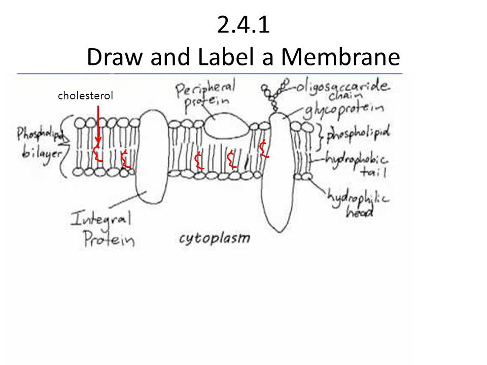
Topic 2.4 MEMBRANES Draw and Label a Membrane cholesterol ...
What is the arrangement of phospholipids in a cell membrane ...
Post a Comment for "39 draw and label a phospholipid bilayer"