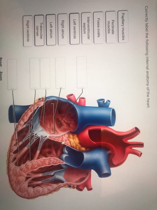
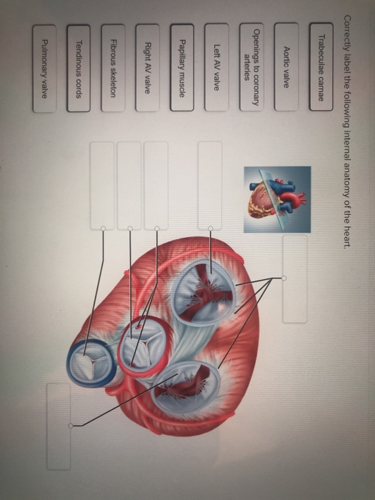
40 correctly label the following internal anatomy of the heart
Heart | Structure, Function, Diagram, Anatomy, & Facts The heart, although a single organ, can be considered as two pumps that propel blood through two different circuits. The right atrium receives venous blood from the head, chest, and arms via the large vein called the superior vena cava and receives blood from the abdomen, pelvic region, and legs via the inferior vena cava. Great Vessels of the Heart: Anatomy & Function - Cleveland Clinic Oct 11, 2022 ... Inferior vena cava. Your great vessels work as a system of highways to keep blood moving in the correct paths throughout your body. These ...
Heart anatomy: Structure, valves, coronary vessels | Kenhub Heart anatomy. The heart has five surfaces: base (posterior), diaphragmatic (inferior), sternocostal (anterior), and left and right pulmonary surfaces. It also has several margins: right, left, superior, and inferior: The right margin is the small section of the right atrium that extends between the superior and inferior vena cava .

Correctly label the following internal anatomy of the heart
Structure of the Heart - SEER Training The internal cavity of the heart is divided into four chambers: Right atrium; Right ventricle; Left atrium; Left ventricle. The two atria are thin-walled ... Chapter 19 Homework Flashcards | Quizlet Correctly label the following internal anatomy of the heart. Drag each label to the location of each structure described. Indicate the heart chamber responsible for the given function. Identify the unique structural characteristics of cardiac muscle. Anatomy & Physiology: The Unity of Form and Function - Quizlet Correctly label the following anatomical features of the heart and thoracic cage. Correctly label the following structures related to the position of the heart in the thorax. Correctly label the following anatomical features of the thoracic cavity. Correctly label the following parts of the pericardium and the heart walls.
Correctly label the following internal anatomy of the heart. Ch. 19 Circulatory System- heart Flashcards | Quizlet Correctly label the following internal anatomy of the heart. Drag each label to the location of each structure described. Explanation The heart functions to first pump deoxygenated blood returning from the body to the lungs in order to release carbon dioxide and reoxygenate the blood. Solved Correctly label the following parts of the internal | Chegg.com Correctly label the following parts of the internal anatomy of the heart. Right pulmonary veins Aorta Left pulmonary veins Pulmonary semilunar Left ventricle Right ventricle Bicuspid valve Tricuspid valve Right pulmonary artery Septum Pulmonary trunk Right atrium Left pulmonary artery Inferior vena cava Left atrium Superior vena cava Reset Zoom Human Heart (Anatomy): Diagram, Function, Chambers, Location in Body The heart is a muscular organ about the size of a fist, located just behind and slightly left of the breastbone. The heart pumps blood through the network of arteries and veins called the... Chapter 22 Heart Flashcards | Quizlet Label the valves in an anterior view of the heart. Label the coronary arteries in an anterior view of the heart. Label the order that blood flows through in the heart, using the arrows as guides. Label the components of the heart wall. Label the components of the heart as seen from a posterior view. Label the major coronary veins.
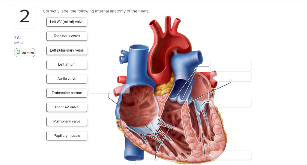
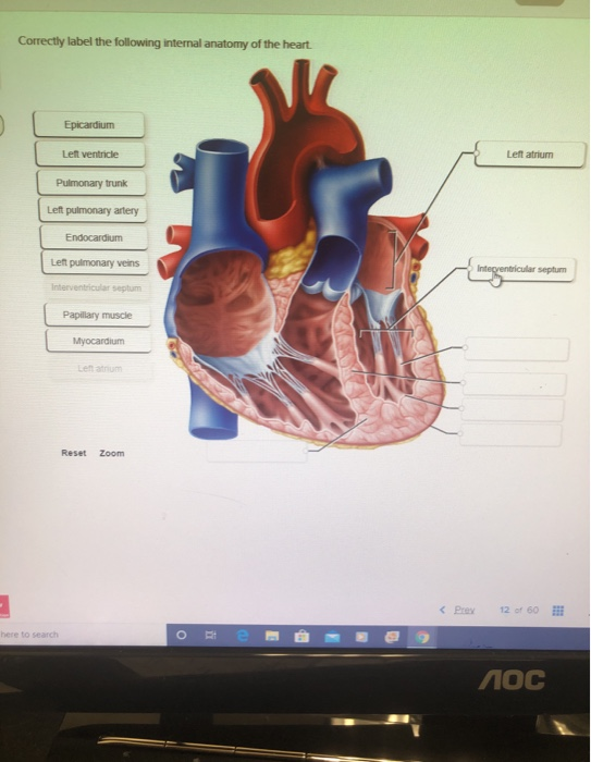
Solved Correctly label the following internal anatomy of the - Chegg Question: Correctly label the following internal anatomy of the heart. Left pulmonary veins Left ventricle Myocardium Endocardium Pulmonary trunk Interventricular septum Left atrium Epicardium Papillary muscle Left pulmonary artery Reset Zoom Show transcribed image text Expert Answer 100% (6 ratings) Starting from Top right to bottom. … A&P II- Exam 1- Heart (Connect) Flashcards | Quizlet Correctly label the following internal anatomy of the heart. Which circuit carries blood from the right ventricle to the lungs for gas exchange and returns it to the left atrium of the heart? Pulmonary Drag each label to the location of each structure described. Correctly label the following internal anatomy of the heart. Chapter 19: The Heart Flashcards | Quizlet The Heart •Circulatory system -heart, blood vessels & blood •Cardiovascular system -heart, arteries, veins and capillaries -2 major divisions •Pulmonary circuit - right side of heart -right heart—lungs—left heart -carries blood to lungs for gas exchange •Systemic circuit - left side of heart -left heart—body—right heart Solved Correctly label the following internal anatomy of the - Chegg Correctly label the following internal anatomy of the heart. 2 Left AV (mitral) valve Tendinous cords 3.84 points 8 00:51:28 Left pulmonary veins Left atrium Aortic valve Trabeculae carnae Right AV valve Pulmonary valve Papillary muscle This problem has been solved!
Label the heart — Science Learning Hub Selecting or hovering over a box will highlight each area in the diagram. In this interactive, you can label parts of the human heart. Drag and drop the text labels onto the boxes next to the heart diagram. If you want to redo an answer, click on the box and the answer will go back to the top so you can move it to another box. AHCDW15Notes17.pdf - 17. Award: 1.00 point Problems? Adjust... Correctly label the following internal anatomy of the heart. Explanation: Blood that has been through the systemic circuit returns by way of the superior and inferior venae cavae to the right atrium. It flows directly from the right atrium, through the right AV valve, into the right ventricle. Solved Correctly label the following parts of the internal - Chegg Answer. Make following changes: In place of right atrium, write left atrium. …. Correctly label the following parts of the internal anatomy of the heart. Place your cursor over the boxes for more information papillary muscles bicuspid valve right atrium septum pulmonary semilunar valve eft atrium chordae tendineae pulmonary semilunar valve ... Heart Diagram with Labels and Detailed Explanation - BYJUS Diagram of Heart. The human heart is the most crucial organ of the human body. It pumps blood from the heart to different parts of the body and back to the heart. The most common heart attack symptoms or warning signs are chest pain, breathlessness, nausea, sweating etc. The diagram of heart is beneficial for Class 10 and 12 and is frequently ...
Pulmonary Vein Isolation With Single Pulse Irreversible ...
Correctly Label The Following Internal Anatomy Of The Heart The aorta, or aortic arch, is the outermost layer of the heart. The left ventricle is covered with the ventricular aorta, and the pulmonary veins are located inside the aorta. The two atria, the left and right aorta, and the right aortic arch are all external organs. These organs carry oxygen-rich blood to the body.
KS2 Heart Diagram QR Labelling Activity - Science - Twinkl
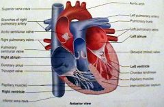
Heart Anatomy: Labeled Diagram, Structures, Blood Flow ... - EZmed Function and anatomy of the heart made easy using labeled diagrams of cardiac structures and blood flow through the atria, ventricles, valves, aorta, pulmonary arteries veins, superior inferior vena cava, and chambers. Includes an exercise, review worksheet, quiz, and model drawing of an anterior vi
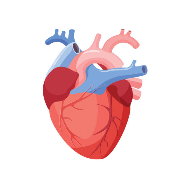
117,700+ Human Heart Stock Photos, Pictures & Royalty-Free ...
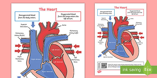
How the Heart Works - What the Heart Looks Like | NHLBI, NIH Heart chambers The two upper chambers of your heart are called atrium, and the two lower chambers are called ventricle. Blood flows from the body and lungs to the atria and from the atria to the ventricles. The ventricles pump blood out of the heart to the lungs and other parts of the body.
Solved Correctly label the following internal anatomy of the ...
Solved Correctly label the following internal anatomy of the - Chegg Correctly label the following internal anatomy of the heart. Openings to coronary arteries Trabeculae carnae Pulmonary valve Tendinous cords Fibrous skeleton Left AV valve Right AV valve Help Tendinous cords Fibrous skeleton Left AV valve Right AV valve Aortic valve Papillary muscle Previous question Next question

Ingestible Sensors and Sensing Systems for Minimally Invasive ...
AHCDW15Notes14 - 14. Award: 1.00 point Problems? Adjust... Correctly label the following internal anatomy of the heart.Explanation:The anterior and posterior interventricular sulci overlie an internal wall, ...
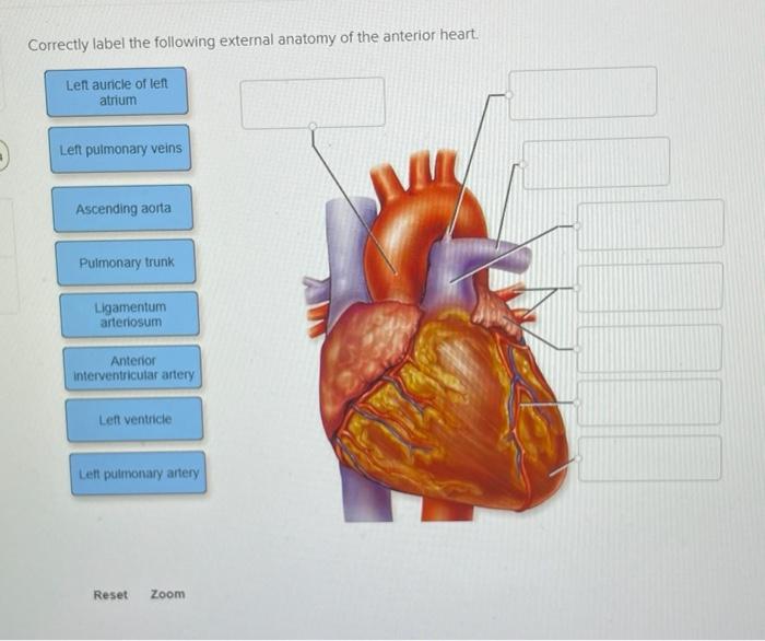
Solved Correctly label the following external anatomy of the ...
Anatomy of the Human Heart - Physiopedia Inform them of the hearts health importance, because it pumps blood and oxygen to all of your organs. When the heart doesn't get the care it needs, ...
Ch17 CV Heart student handout.pptx
Chapter 20-Cardiovascular System Flashcards | Quizlet Correctly label the following internal anatomy of the heart. b Place the labels in order denoting the flow of oxygenated blood through the heart beginning with the vessels that bring blood back to the heart from the lungs. Correctly label the following coronary blood vessels of the heart.
:max_bytes(150000):strip_icc()/human-heart-circulatory-system-598167278-5c48d4d2c9e77c0001a577d4.jpg)
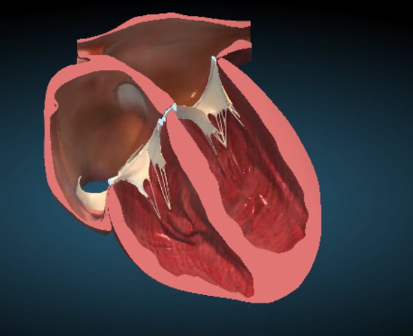
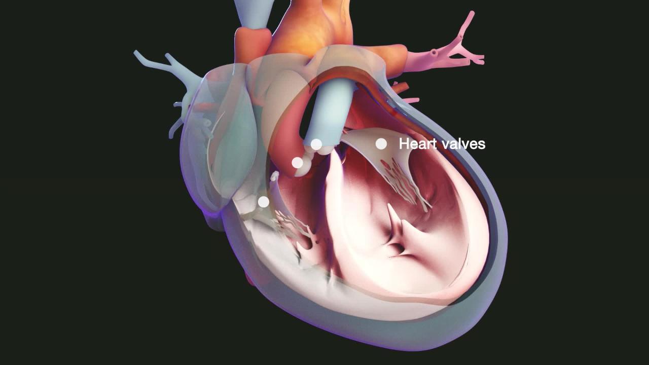
AV and Semilunar Heart Valves
Heart: Anatomy and Function - Cleveland Clinic Your heart is the primary organ of your circulatory system. It pumps blood throughout your body, controls your heart rate and maintains blood pressure. Your heart is a bit like a house. It has walls, rooms, doors, plumbing and an electrical system. All the parts of your heart work together to keep blood flowing and send nutrients to your other ...

Kidney - Wikipedia
Layers of the heart: Epicardium, myocardium, endocardium - Kenhub The myocardium is functionally the main constituent of the heart and the thickest layer of all three heart layers. It is a muscle layer that enables heart contractions. Histologically, the myocardium is comprised of cardiomyocytes.Cardiomyocytes have a single nucleus in the center of the cell, which helps to distinguish them from skeletal muscle cells that have multiple nuclei dispersed in the ...

Chapter 20-Cardiovascular System Flashcards | Quizlet
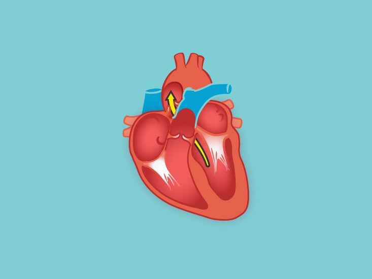
Correctly label the following internal anatomy of the heart. Correctly label the following internal anatomy of the heart. The left atrium, right atrium, and two lower chamber called as the left and right ventricles make up the four chambers that make up the heart. The tricuspid, pulmonary, mitral, and aortic valves are among its four valves. A signal is sent from the SA node to and Av node, which then ...
Heart (right and left atrium): Anatomy and function | Kenhub
Solved Correctly label the following internal anatomy of the - Chegg Question: Correctly label the following internal anatomy of the heart Left atrium A lve Trabeculae camae Le pulmonary veins Pulmonary valve AC Aortic valve Tendinous cords BV Left AV mitral valve Patate Right AV valve Papilary muscle Reset Zoom Show transcribed image text Expert Answer 1st step All steps Answer only Step 1/2

AHCDW15Notes14 - 14. Award: 1.00 point Problems? Adjust ...
The Anatomy of the Heart, Its Structures, and Functions - ThoughtCo The heart is made up of four chambers: Atria: Upper two chambers of the heart. Ventricles: Lower two chambers of the heart. Heart Wall The heart wall consists of three layers: Epicardium: The outer layer of the wall of the heart. Myocardium: The muscular middle layer of the wall of the heart. Endocardium: The inner layer of the heart.

AHCDW15Notes14 - 14. Award: 1.00 point Problems? Adjust ...
The Heart - Anatomy and Physiology II - Lumen Learning In most organs within the body, visceral serous membranes such as the ... Premature removal of these drainage tubes, for example, following cardiac surgery, ...
Solved Correctly label the following internal anatomy of the ...
Solved Correctly label the following internal anatomy of the | Chegg.com Correctly label the following internal anatomy of the heart Right atrium Interventricular septum Right ventricle Right AV (tricuspid) valve Tendinous cords Papillary muscles Fossa ovalis Right atrium Pectinate muscles Right AV (tricuspid) valve This problem has been solved!
Solved Correctly label the following internal anatomy of the ...
CH. 20 Assessment Flashcards | Quizlet Correctly label the following external anatomy of the anterior heart 1. Inferior and superior vena cava 2. Pulmonary trunk 3. Pulmonary veins 4. Aorta 1. right atrium 2. right ventricle 3. left atrium 4. left ventricle Place the steps blood flow through the heart in the correct order. Label the pulmonary and systemic circulation components.

Human Heart Diagram Anatomy Diagram Educational Chart ...
Cardiovascular System Describe the anatomy of the heart. ... Organs. The primary structures that comprise the cardiovascular system: blood vessels ... correct sequence.
Left Ventricle Function, Definition & Anatomy | Body Maps
Solved Correctly label the following external anatomy of the - Chegg Correctly label the following external anatomy of the posterior heart. Left atrium Coronary sinus Left ventricle Right ventricle Apex of heart Left pulmonary veins This problem has been solved! You'll get a detailed solution from a subject matter expert that helps you learn core concepts. See Answer
Fetal Heart – normal – ULTRASOUNDPAEDIA
Anatomy & Physiology: The Unity of Form and Function - Quizlet Correctly label the following anatomical features of the heart and thoracic cage. Correctly label the following structures related to the position of the heart in the thorax. Correctly label the following anatomical features of the thoracic cavity. Correctly label the following parts of the pericardium and the heart walls.
Solved Correctly label the following internal anatomy of the ...
Chapter 19 Homework Flashcards | Quizlet Correctly label the following internal anatomy of the heart. Drag each label to the location of each structure described. Indicate the heart chamber responsible for the given function. Identify the unique structural characteristics of cardiac muscle.

Cells | Free Full-Text | Inherited and Acquired Rhythm ...
Structure of the Heart - SEER Training The internal cavity of the heart is divided into four chambers: Right atrium; Right ventricle; Left atrium; Left ventricle. The two atria are thin-walled ...

ANATOMI PEMBULUH DARAH KEL.1 d4 | PDF

Muscular System Anatomy and Physiology - Nurseslabs

Solved Correctly label the following internal anatomy of the ...

AHCDW15Notes11.pdf - 11. Award: 1.00 point Problems? Adjust ...

Heart: illustrated anatomy | e-Anatomy

AHCDW15Notes15.pdf - 15. Award: 1.00 point Problems? Adjust ...
Conduction system of the heart: Parts and Functions | Kenhub

Chapter 20-Cardiovascular System Flashcards | Quizlet
The Heart

Less than half of adults can correctly label female anatomy ...

Pocket Heart on the App Store
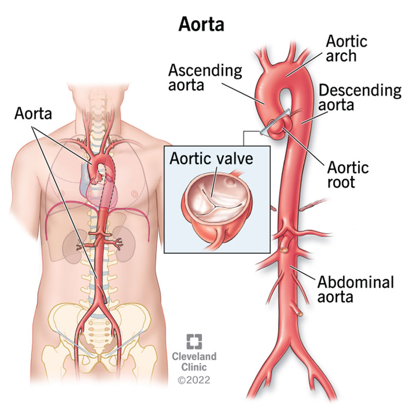
Aorta: Anatomy and Function

Overview of Colic in Horses - Digestive System - MSD ...
Human Anatomy Chapter 1 and 17 Flashcards - Cram.com

Heart Lecture-Exam 3 Flashcards | Quizlet
Respiratory Basics

Heart Anatomy | Anatomy and Physiology II

File:Diagram of the human heart (cropped).svg - Wikipedia

A&P II- Exam 1- Heart (Connect) Flashcards | Quizlet
Structure and Function of the Heart
Post a Comment for "40 correctly label the following internal anatomy of the heart"